Home

частичен чуруликане Comorama cul de sac pleural многомилионен град восък принц

L appareil respiratoire. - PDF Téléchargement Gratuit

IMAGERIE THORACIQUE AUX

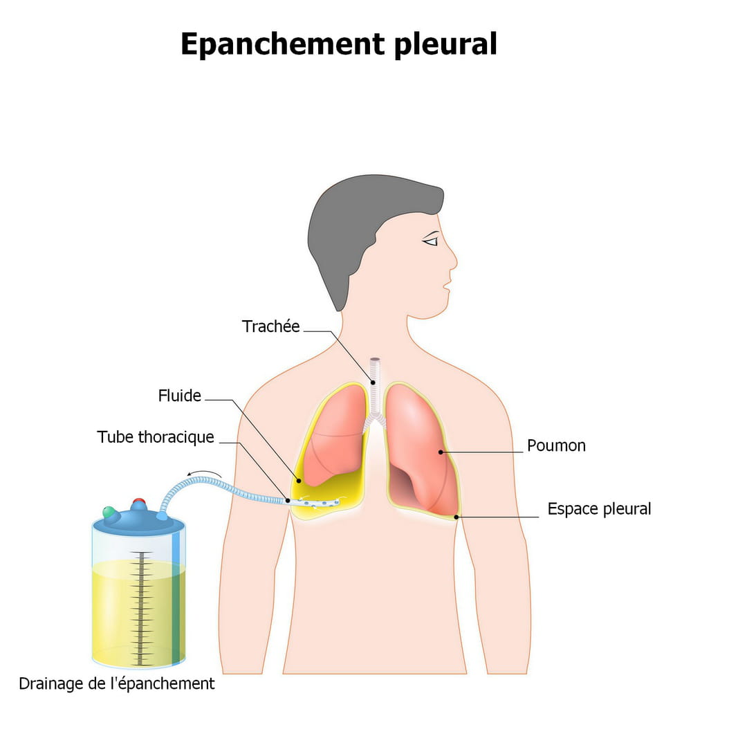
Épanchement pleural : symptômes, causes, complications et ponction

Saga du poumon 12 / . L'épanchement pleural de faible abondance. –  Echo-Urgences
Saga du poumon 12 / . L'épanchement pleural de faible abondance. – Echo-Urgences

InfoCancer - ARCAGY-GINECO - Voies aériennes - Cancer du poumon - Maladie -  Les poumons
InfoCancer - ARCAGY-GINECO - Voies aériennes - Cancer du poumon - Maladie - Les poumons

Hemothorax
Hemothorax

sides:ref-trans:imagerie:multi-items:item_202_199:start [Wiki-SIDES]

Cours
Cours

Semio radio thorax
Semio radio thorax

cul de sac pleural D émoussé -opacité para-médiastinale D
cul de sac pleural D émoussé -opacité para-médiastinale D "en voile latine" : thymus normal pour l'âge -hyperex

Interprétation d'une radiographie thoracique en urgence – 2 – la plèvre |  thoracotomie
Interprétation d'une radiographie thoracique en urgence – 2 – la plèvre | thoracotomie

PPT - Pr Charles-Hugo Marquette PowerPoint Presentation, free download -  ID:6868414
PPT - Pr Charles-Hugo Marquette PowerPoint Presentation, free download - ID:6868414

Interprétation d'une radiographie thoracique en urgence – 2 – la plèvre |  thoracotomie
Interprétation d'une radiographie thoracique en urgence – 2 – la plèvre | thoracotomie

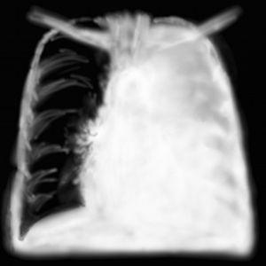
Frontal chest X-ray, left pleural effusion, and blunting of the right... |  Download Scientific Diagram
Frontal chest X-ray, left pleural effusion, and blunting of the right... | Download Scientific Diagram

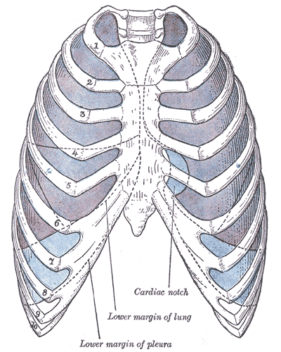
Manual of operative surgery. Fig. 426.—Diagram showing thenormal subpleural  areolar tissue betweenthe pleural reflexion and the diaphragm.{Chevrier^ La  Pr. Med., Jan. 9, 1919.) Fig. 427.—Diagram showing elevationof the pleural  cul-de-sac due
Manual of operative surgery. Fig. 426.—Diagram showing thenormal subpleural areolar tissue betweenthe pleural reflexion and the diaphragm.{Chevrier^ La Pr. Med., Jan. 9, 1919.) Fig. 427.—Diagram showing elevationof the pleural cul-de-sac due

EPANCHEMENT PLEURAL
EPANCHEMENT PLEURAL

312
312

Biotop terminologie médicale, lexique médical, dictionnaire médical, termes  contenant la racine caco, cachexie
Biotop terminologie médicale, lexique médical, dictionnaire médical, termes contenant la racine caco, cachexie

Plèvre — Wikipédia
Plèvre — Wikipédia

Opacity with air fluid level in right basithoracic, associated with... |  Download Scientific Diagram
Opacity with air fluid level in right basithoracic, associated with... | Download Scientific Diagram

UE4 : TD d imagerie n 2. Mascarel. - PDF Téléchargement Gratuit

sides:ref-trans:imagerie:multi-items:item_202_199:start [Wiki-SIDES]

ITEM #202 (ex item 312) - Epanchement Pleural - ppt télécharger

FMPM - Module d'auto apprentissage en pneumologie

Interprétation d'une radiographie thoracique en urgence – 2 – la plèvre |  thoracotomie
Interprétation d'une radiographie thoracique en urgence – 2 – la plèvre | thoracotomie